Bone Cross Section Histology / Biomechanics / In three dimensions an osteon is cylindrical in shape.. Über 7 millionen englischsprachige bücher. Dimitrios mytilinaios md, phd last reviewed: In choosing a technique and processing method, consideration must be given to the type of investigation being carried out. Spongy bone also contains osteocytes housed in lacunae, but they are not arranged in concentric circles. Bone cross section slide labeled :
However, this does not preserve the cells very well. The darker ring consists of layers of bone matrix made by cells called osteoblasts Histology spinal cord and ganglion : There are a number of options available when the histologist is required to produce sections from bone or other calcified specimens. Acid or chelators are used to remove minerals from the matrix and decalcify the bone.
Bone Histology - Biology bibliographies - Cite This For Me from mesa-anatomy.weebly.com In addition to discussing the cellular constituents of bone and the architectural arrangement of their products, this article will also address the embryology and mechanisms of ossification as well. To the left is muscle tissue, and to the right is bone marrow. The matrix is about 50% inorganic matter, with calcium and phosphorus forming hydroxyapatite crystals. They offer a reasonably consistent picture of their structural pattern. The red arrow indicates a haversian canal; A long bone has two parts: Be able to recognize these cell types: Later discussions in this chapter will show that bone is also dynamic in that its shape adjusts to accommodate stresses.
Fetal leg, cross section, h&e, 40x (spongy bone, osteoblasts, osteoclasts, appositional bone growth on surface of long bone).
Spongy bone, also known as cancellous bone or trabecular bone, looks like a sponge under the microscope. The red arrow indicates a haversian canal; This photo shows a cross section through bone. The basic unit of structure in this type of bone is the haversian system, or osteon. This section will examine the gross anatomy of bone first and then move on to its histology. Über 7 millionen englischsprachige bücher. *none of the slide images above are shown at their actual scale. About press copyright contact us creators advertise developers terms privacy policy & safety how youtube works test new features press copyright contact us creators. Gross anatomy of bone the structure of a long bone allows for the best visualization of all of the parts of a bone (figure 6.7). Hematoxylin eosin stain (human) 1.6 blood smear: Окраска по методу шморляпластинчатая (тонковолокнистая) костная ткань в. 7 minutes bone formation in a developing embryo begins in mesenchyme and occurs through one of two processes: June 17, 2021 reading time:
(teased tendon) 1.9 elastic tissue: Hematoxylin eosin stain (human) 1.6 blood smear: A thorough knowledge of the two imaging … In three dimensions an osteon is cylindrical in shape. Later discussions in this chapter will show that bone is also dynamic in that its shape adjusts to accommodate stresses.
Shotgun Histology Dense Bone - YouTube from i.ytimg.com The inner portion of the bone is composed of trabecular bone and the intervening bone marrow. Bone cross section slide labeled : Gross anatomy of bone the structure of a long bone allows for the best visualization of all of the parts of a bone (figure 6.7). Decalcified section of compact bone, cross section, 150x. June 17, 2021 reading time: Be able to describe, as well as recognize in microscope sections/photos, the process of intramembranous bone formation, including the process by which cancellous bone is converted into compact bone. Each osteon looks like a ring with a light spot in the center. There are a number of openings and canals in the temporal bone through which structures enter and exit the cranial cavity.
This model shows a cross section of compact bone.
Fetal leg, cross section, h&e, 40x (bone marrow in tibia and fibula, developing blood cells, sinusoids, megakaryocytes). This section will examine the gross anatomy of bone first and then move on to its histology. At the outer regions of the section, you can see a dense, thick layer of compact bone. Gross anatomy of bone the structure of a long bone allows for the best visualization of all of the parts of a bone (figure 6.7). This section will examine the gross anatomy of bone first and then move on to its histology. Bone matrix and cells bone matrix osseous tissue is a connective tissue and like all connective tissues contains relatively few cells and large amounts of extracellular matrix. Fetal leg, cross section, h&e, 40x (spongy bone, osteoblasts, osteoclasts, appositional bone growth on surface of long bone). Bone is hard and many of its functions depend on that characteristic hardness. While it is not as hard as compact bone, spongy bone plays an important role of protecting the marrow where blood cells are produced. Either endochondral or intramembranous osteogenesis (ossification).intramembranous ossification is characterized by the formation of bone. Cross section (human) 1.5 blood smear: There are a number of options available when the histologist is required to produce sections from bone or other calcified specimens. Bianca fiorentino slotfeldt changed description of no.
In addition to discussing the cellular constituents of bone and the architectural arrangement of their products, this article will also address the embryology and mechanisms of ossification as well. Transverse cross section of compact bone tissue; Concentric layers of bone cells (osteocytes) and bone matrix surround the central canal. Histology spinal cord and ganglion : To the left is muscle tissue, and to the right is bone marrow.
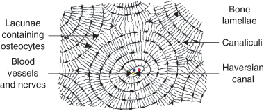
1 Oral embryology, histology and anatomy | Pocket Dentistry from pocketdentistry.com 7 minutes bone formation in a developing embryo begins in mesenchyme and occurs through one of two processes: Bone tissue (osteons or haversian systems). In the center of each osteon is the central canal, a space that houses blood vessels and nerves that supply bone. Decalcified section of compact bone, cross section, 150x. In addition to discussing the cellular constituents of bone and the architectural arrangement of their products, this article will also address the embryology and mechanisms of ossification as well. The basic unit of structure in this type of bone is the haversian system, or osteon. Über 7 millionen englischsprachige bücher. Gross anatomy of bone the structure of a long bone allows for the best visualization of all of the parts of a bone (figure 6.7).
The choice of ct versus mr depends on the structures and the disease processes that require assessment, delineation, and characterization.
To the left is muscle tissue, and to the right is bone marrow. Each osteon looks like a ring with a light spot in the center. In three dimensions an osteon is cylindrical in shape. Be able to recognize these cell types: Bone tissue (osteons or haversian systems). Very inneficient way to merge verticles. The red arrow indicates a haversian canal; The basic unit of structure in this type of bone is the haversian system, or osteon. Be able to describe, as well as recognize in microscope sections/photos, the process of intramembranous bone formation, including the process by which cancellous bone is converted into compact bone. There are a number of openings and canals in the temporal bone through which structures enter and exit the cranial cavity. Gross anatomy of bone the structure of a long bone allows for the best visualization of all of the parts of a bone (figure 6.7). Long section (tendon) 1.8 white fibrous connective tissue: This slide contained a cross section of a very small bone, and you are looking at the entire thickness of the shaft of the bone.
Hematoxylin eosin stain (human) 16 blood smear: bone cross section. The osteocytes are arranged in concentric rings of bone matrix called lamellae (little plates), and their processes run in interconnecting canaliculi.


0 Komentar